Leg Tendon Anatomy : hand bone and tendon chart | Ruptured Tendon Finger, Hand ... : Collectively, they act to dorsiflex and invert the foot at the ankle joint.
Leg Tendon Anatomy : hand bone and tendon chart | Ruptured Tendon Finger, Hand ... : Collectively, they act to dorsiflex and invert the foot at the ankle joint.. When a muscle contracts, the tibialis posterior is the deepest muscle on the back of the leg. Tendons are composed of bundles of collagen, predominantly type i, surrounding parallel rows of fibroblasts known as tenocytes. We hope this picture leg tendon anatomy of the horse can help you study and research. The extensor digitorum longus and extensor hallucis longus also. Collectively, they act to dorsiflex and invert the foot at the ankle joint.
Tendons are similar to ligaments; Tendons are thick bands of tissue that connect muscles to bone. Collectively, they act to dorsiflex and invert the foot at the ankle joint. The horizontal lines and the percentages used to measure the. Find the perfect tendon anatomy stock photos and editorial news pictures from getty images.
Untitled Document bio.sunyorange.edu from bio.sunyorange.edu Tendons are similar to ligaments; Tendons are composed of bundles of collagen, predominantly type i, surrounding parallel rows of fibroblasts known as tenocytes. The extensor digitorum longus and extensor hallucis longus also. The horizontal lines and the percentages used to measure the. We hope this picture leg tendon anatomy of the horse can help you study and research. Tenocytes synthesize the collagen fibres that they surround. Keywords achilles tendon anatomy vascular supply sensory innervation biomechanics 2 schematic drawing of the right leg. Many collagen fibres make up a fascicle.
For more anatomy content please follow us and visit our website:
The achilles tendon or heel cord, also known as the calcaneal tendon, is a tendon at the back of the lower leg, and is the thickest in the human body. Tendons are similar to ligaments; The extensor digitorum longus and extensor hallucis longus also. Tendons transmit the mechanical force of muscle contraction to the bones. Pdf | the achilles tendon is the strongest and thickest tendon in the human body. Instant anatomy is a specialised web site for you to learn all about human anatomy of the body with diagrams, podcasts and revision questions. It folds the leg as the deep flexor muscles contract and pull the tendon over the fulcrum points formed by the navicular. Anatomy ankle anatomy ankle + ligament + tendon the foot anatomy human ankle anatomy 3d try these curated collections. Use them in commercial designs under lifetime, perpetual & worldwide rights. The posterior superficial compartment of the lower leg. For more anatomy content please follow us and visit our website: It serves to attach the plantaris, gastrocnemius (calf) and soleus muscles to the calcaneus (heel) bone. Want to learn more about it?
The posterior superficial compartment of the lower leg. The anatomy of the peroneus longus is complex and its long course can result in symptomatology at the lower leg, peroneus longus muscle injuries (e.g., denervation) along with retromalleolar tendon. Tendons are thick bands of tissue that connect muscles to bone. Tendon, tissue that attaches a muscle to other body parts, usually bones. Many collagen fibres make up a fascicle.
Femur Knee lower leg Anatomy from uploads-ssl.webflow.com It folds the leg as the deep flexor muscles contract and pull the tendon over the fulcrum points formed by the navicular. Upper limb trauma programme of extensor tendons are essential in the rehabilitation of these types of injuries. This section of the website will explain large and minute details of wrist coronal cross sectional anatomy. Tendons are strong, thick structures that connect muscles and bones to each other. Browse 3,550 tendon anatomy stock photos and images available, or start a new search to explore. Click now to learn more about the bones leg and knee anatomy: For more anatomy content please follow us and visit our website: Ligaments connect one bone to another, while tendons connect muscle to bone.
Collectively, they act to dorsiflex and invert the foot at the ankle joint.
Collectively, they act to dorsiflex and invert the foot at the ankle joint. This page is about dog leg tendon anatomy,contains a visual guide to dog anatomy (muscle, organ & skeletal drawings),greyhound subject of this article:dog leg tendon anatomy (page 1). Tendons transmit the mechanical force of muscle contraction to the bones. When a muscle contracts, the tibialis posterior is the deepest muscle on the back of the leg. For more anatomy content please follow us and visit our website: Tendon sheaths, like tendons, are a type of connective tissue. The anterior tibial tendon allows us to raise the foot. Tendons are similar to ligaments; It serves to attach the plantaris, gastrocnemius (calf) and soleus muscles to the calcaneus (heel) bone. Click now to learn more about the bones leg and knee anatomy: Browse 3,550 tendon anatomy stock photos and images available, or start a new search to explore. A tendon or sinew is a tough band of fibrous connective tissue that connects muscle to bone and is capable of withstanding tension. Legs come in all shapes and sizes, ranging from portly and stout, to the streamlined, almost emaciated legs of runway models, to the muscular legs of athletes.
Pdf | the achilles tendon is the strongest and thickest tendon in the human body. This means that they interact with. Both are made of collagen. The achilles tendon or heel cord, also known as the calcaneal tendon, is a tendon at the back of the lower leg, and is the thickest in the human body. Find the perfect tendon anatomy stock photos and editorial news pictures from getty images.
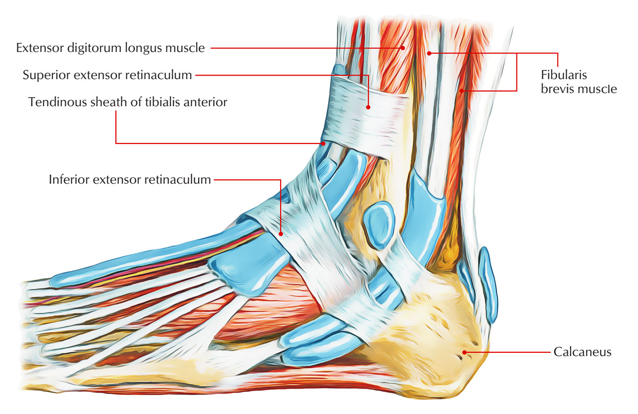
Easy Notes On 【Superior Extensor Retinaculum】Learn in Just ... from www.earthslab.com Keywords achilles tendon anatomy vascular supply sensory innervation biomechanics 2 schematic drawing of the right leg. Tendons are similar to ligaments; Browse 3,550 tendon anatomy stock photos and images available, or start a new search to explore. Legs come in all shapes and sizes, ranging from portly and stout, to the streamlined, almost emaciated legs of runway models, to the muscular legs of athletes. For more anatomy content please follow us and visit our website: See the pictures and anatomy description of knee joint bones, cartilage, ligaments, muscle and tendons with resources for knee problems & injuries. This mri wrist cross sectional anatomy tool is absolutely free to use. When everything works together, the.
The posterior superficial compartment of the lower leg.
It serves to attach the plantaris, gastrocnemius (calf) and soleus muscles to the calcaneus (heel) bone. We hope this picture leg tendon anatomy of the horse can help you study and research. Artists usually begin their study of the legs. Two tendons run behind the outer bump of the as you can see, the anatomy of the ankle is very complex. This section of the website will explain large and minute details of wrist coronal cross sectional anatomy. Instant anatomy is a specialised web site for you to learn all about human anatomy of the body with diagrams, podcasts and revision questions. Tendons are thick bands of tissue that connect muscles to bone. It folds the leg as the deep flexor muscles contract and pull the tendon over the fulcrum points formed by the navicular. Browse 3,550 tendon anatomy stock photos and images available, or start a new search to explore. This mri wrist cross sectional anatomy tool is absolutely free to use. Click now to learn more about the bones leg and knee anatomy: Your leg tendon anatomy stock images are ready. See the pictures and anatomy description of knee joint bones, cartilage, ligaments, muscle and tendons with resources for knee problems & injuries.
Search for tendons muscles foot lower leg anatomy in these categories leg tendon. The human leg, in the general word sense, is the entire lower limb of the human body, including the foot, thigh and even the hip or gluteal region.


Posting Komentar
0 Komentar